
Study reveals how cancer cells metastasise to the liver
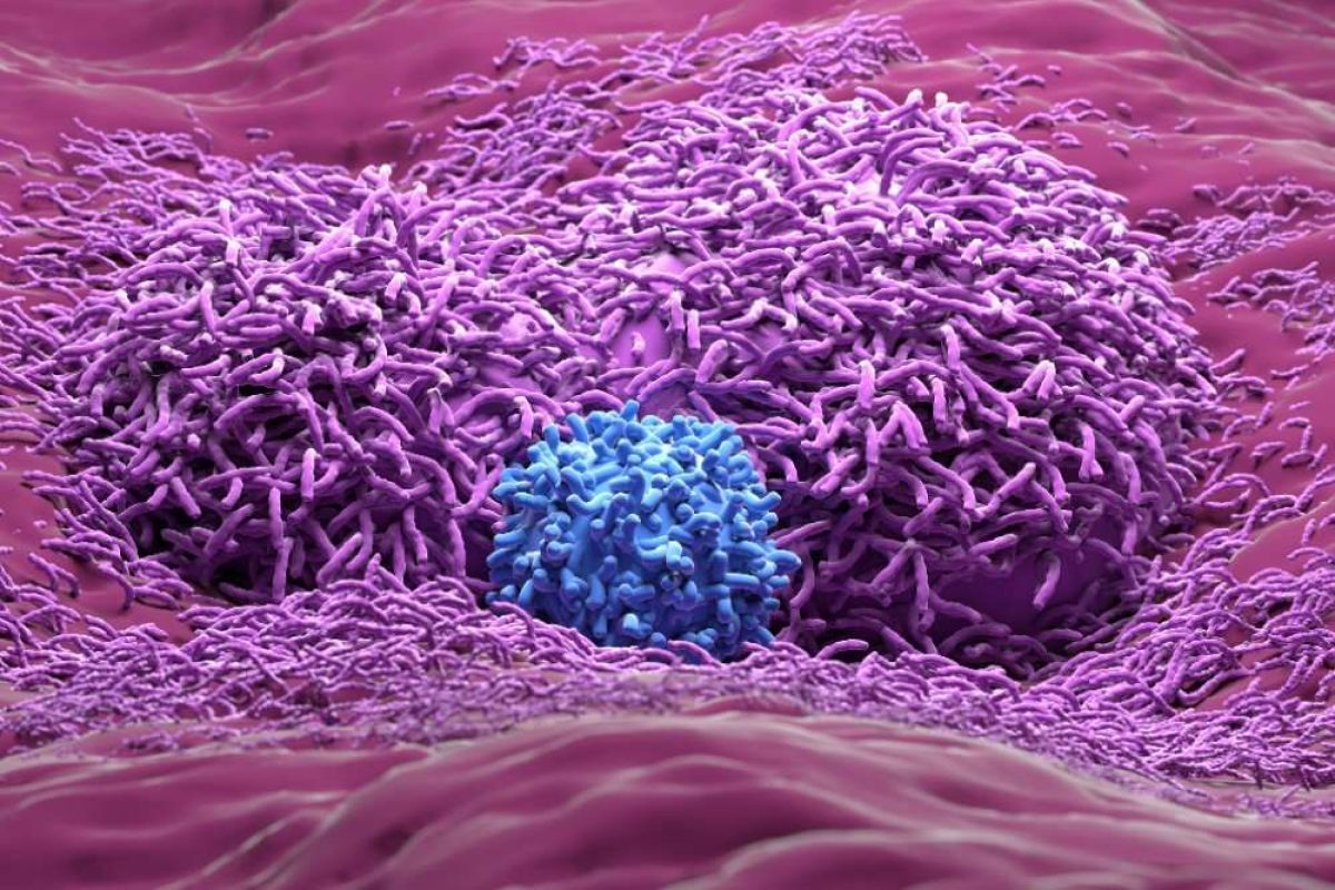
Researchers at ETH Zurich in Switzerland have uncovered how colorectal cancer cells colonise the liver.
The findings, by Andreas Moor in the Department of Biosystems Science and Engineering and his team, will help to develop treatments with which they hope could hinder the metastatic process.
Cancer metastasises when cells from the primary tumour break off and travel via the circulatory system to other parts of the body. For colorectal cancer cells, the liver is the last stop and they get caught in the liver’s capillary network.
Moor’s team have now shown that the liver cells also play a large role in whether the cancer cells lodged there can colonise their new location. When liver cells have a protein called Plexin-B2 and the colorectal cancer cells possess certain proteins from the semaphorin family, the colorectal cancer cells can attach themselves to the liver cells.
Cancer cells that have semaphorins on their surface are especially dangerous. The study data show that colorectal cancer metastasises earlier and more frequently to the liver if the tumour has large amounts of semaphorin.
Moor and his team discovered that direct contact between plexin and semaphorin triggers fundamental changes in colorectal cancer cells. To break off from the primary tumour, the cancer cells have to change their identity: they free themselves from the surface layer of the intestine, or epithelium, severing their close connections to neighbouring cells.
Once in the bloodstream, the cancer cells resemble those from connective tissue called mesenchyme. Yet once they find their new niche – thanks to the plexin on some liver cells – the cancer cells turn back into their sedentary form.
Further tests have shown that plexin also encourages the formation of metastases in melanoma and pancreatic cancer, suggesting new avenues of research.
Diana Spencer, Senior Digital Content Editor, DDW