
Nanobody-based 3D imaging boosts neuroscience research
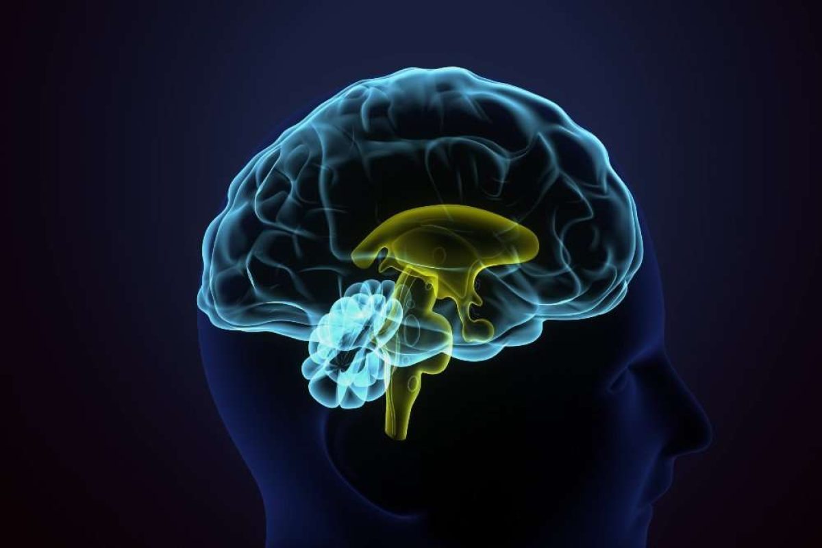
Scientists have created a nanobody-based method that enables ultra-fast, high-resolution 3D mapping of neurons and glia in thick brain tissues.
The researchers from Japan have developed a fast, high-sensitivity three dimensional immunohistochemistry (3D-IHC) method using nanobodies fused with peroxidase and a novel signal amplification system.
Their technique labels neurons and glia in 1mm-thick brain tissue within three days, offering a powerful new tool for neuroscience and disease research, including Alzheimer’s pathology.
Traditional 3D-IHC methods are often time-consuming and suffer from poor antibody penetration, which limits their effectiveness in deep tissues.
The new 3D-IHC technique, developed by Professor Kenta Yamauchi and Professor Hiroyuki Hioki of Juntendo University in Tokyo, dramatically improves the speed, depth, and sensitivity of immunolabeling in thick tissue samples.
The study published in Communications Biology employs a method called POD-nAb/FT-GO 3D-IHC, which combines nanobodies fused with peroxidase (POD-nAbs) and a fluorescent signal amplification system known as Fluorochromized Tyramide-Glucose Oxidase (FT-GO).
The technique also proved effective in a disease model. In brain slices from Alzheimer’s disease model mice, the method revealed activated microglia clustering around beta-amyloid plaques, an important hallmark of neuroinflammation and disease progression.
Compared to standard fluorescent protein imaging and direct labelling with synthetic fluorophore-conjugated nanobodies, the POD-nAb/FT-GO protocol provided up to nine-fold greater signal intensity at tissue depths of 500 microns.
This substantial signal amplification reduces the need for prolonged imaging sessions, making the method particularly suited for high-throughput analysis.
The method has some limitations, for example, the authors note that signal homogeneity decreases in tissues thicker than 1 mm. Additionally, quantitative comparisons of antigen expression are challenging due to the non-linear nature of enzymatic signal amplification.
Another hurdle is the limited availability of nanobodies suitable for histochemical labelling, though this is expected to improve as more nanobody sequences become publicly available.
Diana Spencer, Senior Digital Content Editor, DDW