
Vaccines trigger rapid lymph node responses, study finds
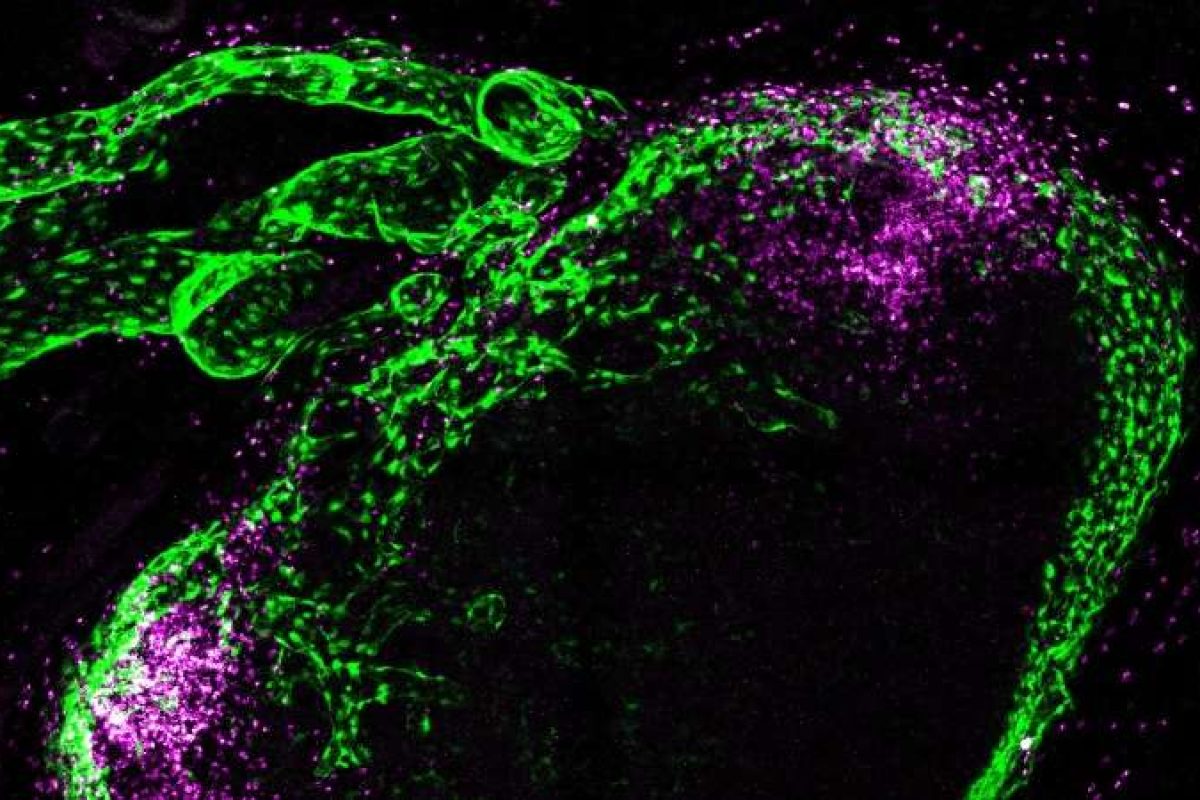
Vaccines trigger a notably rapid response in the stromal cells of draining lymph nodes within the first hours after administration.
Researchers at the University of Turku and the InFLAMES Flagship in Finland also discovered that the stromal alterations prime the lymph node landscape for the subsequent steps of vaccine-induced immune responses.
Researchers observed that lymphatic endothelial cells and other stromal cells are the first cells in the lymph nodes to come into contact with vaccines.
The vaccines induced several changes in stromal cells at the gene and protein levels within the first hours after vaccination, which in turn affected lymph node function.
The researchers also discovered that different vaccines activate lymph node stromal cells in different ways.
“The general perception is that specialised white blood cells begin to transport the active ingredients of a vaccine from the muscle to the lymph nodes approximately 12 hours after vaccination. Our new research results show that the vaccine-induced immune response of the lymph nodes begins much earlier, and the white blood cells bearing the antigens, or the active ingredients of the vaccine, actually arrive at an altered tissue environment that has already been primed by the vaccine. This priming supports the development of the immune response,” explains Postdoctoral Researcher of the Research Council of Finland Ruth Fair-Mäkelä, the lead author of the study.
The study has been published in the journal Science Immunology.
InFLAMES Flagship is a joint initiative of the University of Turku and Åbo Akademi University in Turku.
Diana Turner, Senior Digital Content Editor, DDW
Image: The image is from a draining lymph node (approximately 2mm in size) after vaccination. The green cells are stromal cells that line the lymph node sinuses. The lymphatic vessels of the lymph node are visible at the top left. The pink dots are eosinophilic white blood cells that have accumulated in clusters within the lymph node. There are also eosinophils in the adipose tissue surrounding the lymph nodes. © Pinja Thorén.