
Biomolecular condensates finding could shape drug development
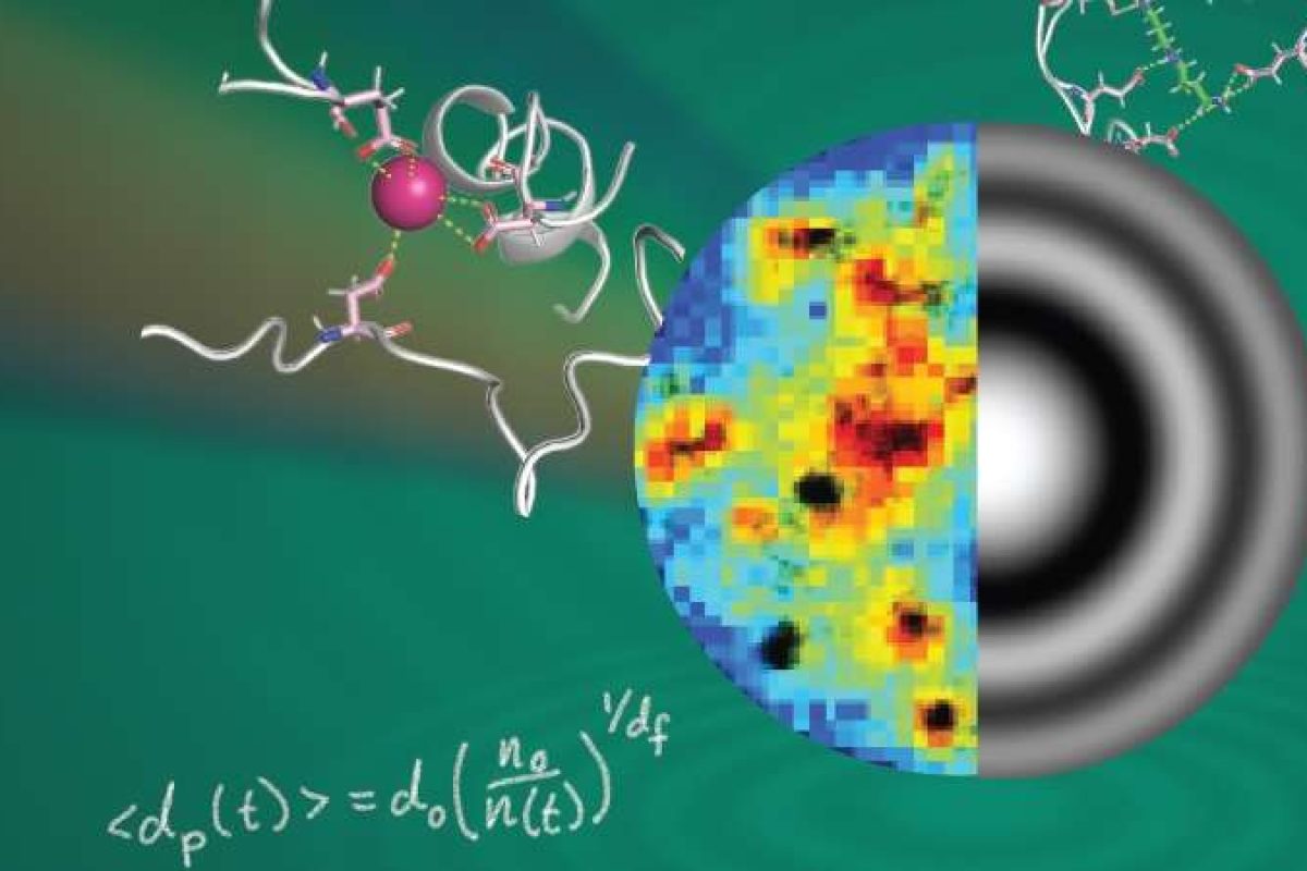
A team of New York University (NYU) chemists and physicists have unlocked how cells build and grow tiny, dynamic droplets known as biomolecular condensates.
For the first time, scientists measured the protein content and growth dynamics of individual biomolecular condensates without disturbing them, gaining insights that may shape future drug development and disease modelling.
Biomolecular condensates manage vital cellular functions, from regulating genes to responding to stress. Until now, studying them has involved distorting them.
“It’s been the elephant in the room for scientists,” said Saumya Saurabh, Assistant Professor of Chemical Biology at NYU and the senior author of the new study, published in the Journal of the American Chemical Society. “Our research provides a precise and non-invasive way to study biomolecular condensates.”
“Being able to see ‘under the hood’ for the first time has revealed some big surprises about this important class of systems,” added study author David Grier, Professor of Physics and Director of the Center for Soft Matter Research at NYU.
Innovative imaging techniques
To study biomolecular condensates under the microscope, researchers have traditionally been limited to using fluorescent tags or two-dimensional surfaces, both of which can significantly disturb the droplets’ behaviour.
Grier’s lab has pioneered the use of holographic microscopy, which uses lasers and lenses to create three-dimensional images, or holograms, of particles that are captured on video for analysis.
The technique allows scientists to flow particles in a solution so that they can be clearly seen and individually characterised—without the need for fluorescent labels or attachment to a surface. They found that relevant biomolecules could concentrate proteins more than ten-fold inside condensates.
The team then utilised super-resolution imaging, a Nobel Prize-winning technology, which revealed that condensates were not simple uniform droplets but exhibited intricate nanoscale organisation, a realm 1,000 to 100,000 times smaller than the width of a human hair. The findings were supported by molecular dynamics simulations.
The authors say that understanding how biomolecular condensates are organised and grow may hold clues for treating a range of illnesses, from cancer and infectious diseases to neurological disorders.
In addition, scientists recently discovered that many drug molecules end up inside biomolecular condensates in the cells. This sequestering of drugs within condensates may help explain why drugs that are made to target a specific protein still cause side effects.
Diana Turner, Senior Digital Content Editor, DDW
Image: Researchers peer inside biomolecular condensates—tiny, membraneless compartments within cells—using super-resolution microscopy (colorful visualization on left) and holographic microscopy (represented by concentric rings on right) to unlock the complex secrets of these essential cellular structures. Credits: Julian von Hofe and Saumya Saurabh, PhD.